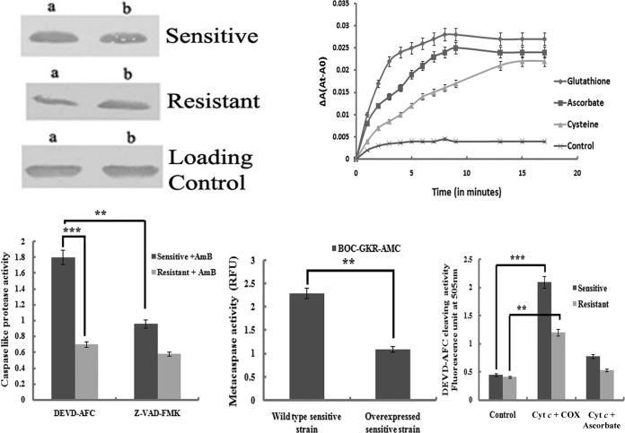
FIG 7.
(Top left) Western blot analysis to detect the release of cytochrome c into the cytoplasm of differentially treated cells of AmB-sensitive and -resistant strains. Lanes a, cells treated with AmB (0.125 μg/ml) for 24 h; lanes b, cells preincubated with aminotriazole and then AmB for 24 h. (Top right) Kinetic profiles of Cyt c reduction by various thiols and ascorbate. ΔA(At-A0), change in absorbance times the absorbance at the indicated time minus the absorbance at time zero. (Bottom left) Comparative study of AmB-induced activation of caspase 3-like protease in sensitive and resistant promastigotes and its downstream effects in the presence of the caspase 3 inhibitor. (Bottom center) Metacaspase activities in the sensitive wild type and in the sensitive strain in which LdAPx was overexpressed. (Bottom right) Oxidized cytochrome c (COX) rapidly activated caspase 3 in a cytosolic fraction of the sensitive strain, unlike in the resistant strain, whereas cytochrome c reduced by ascorbate or cysteine or glutathione was completely ineffective at activating the caspases in the case of the resistant strain. **, significant difference (P < 0.05); ***, significant difference (P < 0.005).

