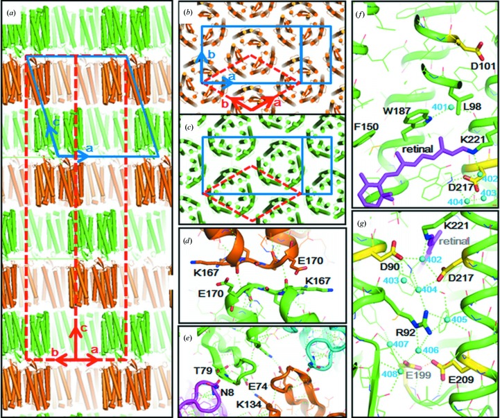
Figure 2.
(a, b, c) Protein packing in the hexagonal crystal of aR2 used in this study. The protein arrangement is described by space group H32 (red broken lines) or C2 (solid blue lines). (d) Intermembrane protein–protein contacts on the cytoplasmic side. (e) Intermembrane protein–protein contacts on the cytoplasmic surface. (f, g) Distribution of water molecules (cyan spheres) in proton-uptake and proton-release channels.

