Figure 3.
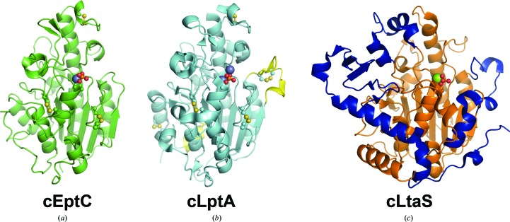
Overall structures of the pEtN transferases cEptC and cLptA and the B. subtilis lipoteichoic acid synthase cLtaS. (a) cEptC (green), with phosphothreonine and disulfides shown in ball-and-stick representation and the Zn2+ ion as a purple sphere. (b) cLptA (PDB entry 4kay; cyan), with phosphothreonine and disulfides shown in ball-and-stick representation and Zn2+ ions as purple spheres. Structural insertions found in cLptA, but not cEptC, are colored yellow. cLptA and cEptC superpose with a Cα r.m.s.d. of 0.59 Å over 211 residues. (c) B. subtilis cLtaS (PDB entry 2w8d; orange), with phosphothreonine shown in ball-and-stick representation and the Mg2+ ion as a green sphere. Structural features unique to cLtaS are colored blue. cLtaS and cEptC superpose with a Cα r.m.s.d. of 3.74 Å over 216 residues.

