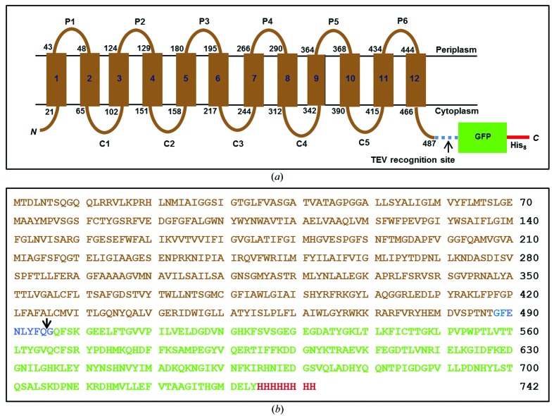
Figure 1.
Schematic representation of the LysP-GFP8His fusion construct. (a) The topology of LysP predicted by the TMHMM online server (http://www.cbs.dtu.dk/services/TMHMM-2.0/) shows 12 transmembrane helices (numbered rectangles). The start and end residue numbers for each transmembrane helix are shown at the membrane boundary (black lines). P1–P6 and C1–C5 designate the periplasmic and cytoplasmic loops, respectively. The C-terminal GFP8His fused to LysP via a linker that has a TEV protease recognition site (blue dashed line) is shown as a green box with the extension as a red line. The Cin topology ensures that the GFP resides in the reducing environment of the cytoplasm, which is necessary for proper folding of this β-barrel reporter. (b) The deduced amino-acid sequence of the LysP-GFP8His construct. Sequences corresponding to LysP, the TEV protease-containing linker, the GFP and the His tag are shown in brown, blue, green and red, respectively. The arrow marks the TEV protease cut site.

