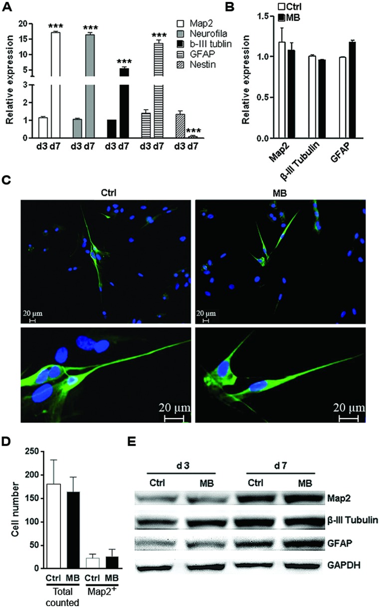
FIGURE 4.
MB does not impede committed neuronal differentiation. (A) Q-RTPCR analysis of the expression of differentiation markers in differentiated NPCs at 1 and 7 days after growth factors withdraw. N = 3 per group. ***p < 0.001 compared with day-1 differentiated NPCs. (B) Q-RTPCR for expression of differentiation markers in differentiated NPCs in the presence or absence of MB. N = 3 per group. (C) Map2 staining for differentiated NPCs in the presence or absence of MB. Upper, 50×; lower, 200×. (D) Total cell number counting and Map2+ cell number in five microscopy fields (50×). (E) Western blot assay for expression of differentiation markers in differentiated NPCs in the presence or absence of MB. This is a representative of two independent experiments.

