FIGURE 1.
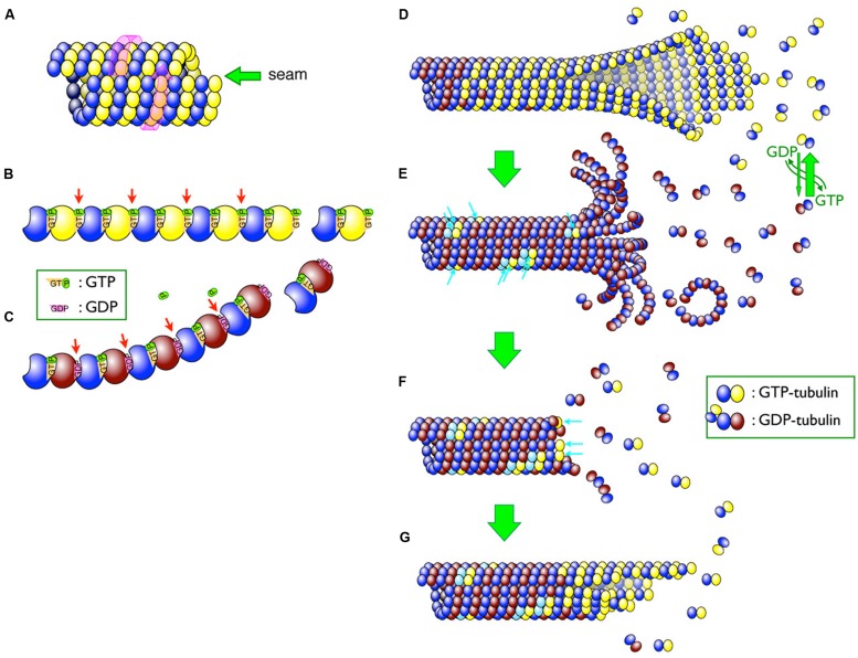
Schematic diagram of microtubules. (A) Lateral arrangement of tubulin dimers in the microtubule. The tubulin molecules are laterally aligned along the three-start left-handed helix (indicated by a pink wrap-around arrow) and different tubulin molecules thus meet at a seam (indicated by a green arrow). (B,C) GTP hydrolysis causes a conformational change in protofilaments. Whereas GTP–tubulins (B) have a straight conformation that fits well in the wall of the microtubule, GDP-tubulins (C) tend to curve outward from the microtubule lattice. β-tubulins bound to GDP are shaded brown, whereas those bound to GTP are yellow. Changes in inter-dimer interactions are indicated by arrows. (D–G) Depolymerization and rescue of microtubules. While newly incorporated tubulin dimers are all GTP–tubulins (D), GTP molecules are relatively quickly hydrolyzed and most of the dimers in the microtubule become GDP-tubulin (shaded in brown; D) Remnant GTP–tubulins in the microtubule are indicated by blue arrows (E). Depolymerization can be stopped by remnant GTP–tubulins as a “rescue” action (F). The addition of new GTP-tubulins (blue arrows in F) resumes the growth, and the addition of the tubulin dimers continues (G).

