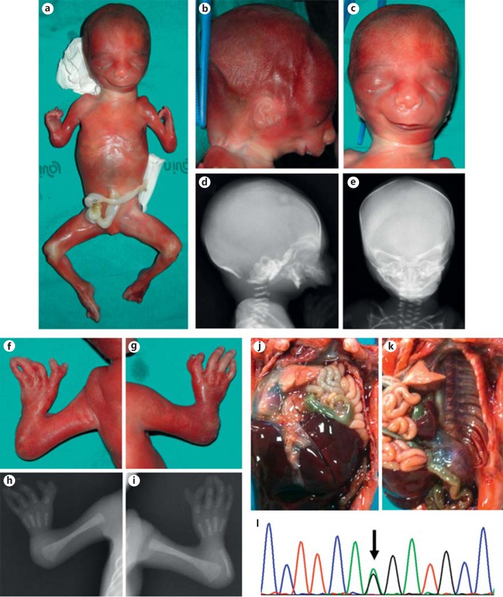
Fig. 1.
a Whole-body picture of the fetus showing typical appendicular and craniofacial features. b-e Comparison of the clinical (b right lateral view, c frontal view) and radiographic (d right lateral view, e frontal view) presentation of the craniofacial anomalies. f-i Comparison of the clinical (f left hand, g right hand) and radiographic (h left hand, i right hand) presentation of the hand anomalies. j Cranial dislocation of the gut through a large defect of the left diaphragm. k Partial evisceration showing complete agenesis of the left hemi-diaphragm. l Direct sequencing electropherogram of the acceptor splicing junction of SF3B4 exon 2, showing the heterozygous mutation c.35-2A>G.

