
Figure 6.
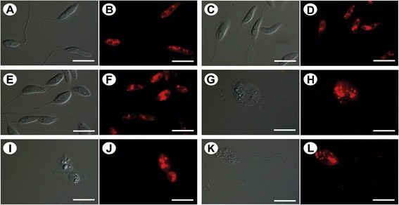
Differential interference contrast microscopy (DIC) and fluorescence microscopy with Nile Red staining of Leishmania amazonensis promastigotes without treatment (A, B) and treated with concentrations that corresponded to the IC 50 (C-F) and IC 90 (G-L) of BZTS for 48 h. In the treated promastigotes, the images suggest the concentration-dependent accumulation of lipid-storage bodies in the cytoplasm. (G, I, K) The parasites that were treated with the IC90 of BZTS were completely modified. Scale bar = 10 μm.