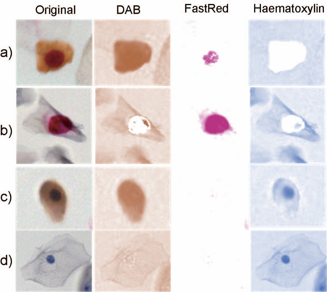
Figure 3.
Color deconvolution in images of epithelial cells with different staining patterns: a) p16+/Ki67+, b) p16−/Ki67+, c) p16+/Ki67−, and d) p16−/Ki67− . Respective columns labeled DAB and FastRed display localization of individual stains after deconvolution demonstrating that only FastRed and Haematoxylin images are suitable for nuclear segmentation.

