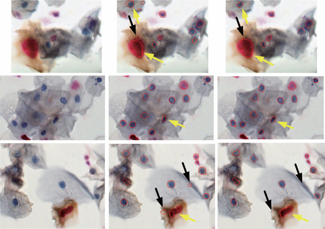
Figure 6.
Example segmentations of nuclei in Pap smears stained for Ki67 and p16. Boundaries of automatically delineated nuclei are marked in red. Left column - original images, middle column - results from RSD – our previously developed algorithm, and right column – final output of the proposed method involving RSD combined with superpixel analysis. Yellow arrows (in the right column) show contour detection improvement after implementation of superpixels. Black arrows point onto areas detected by the RSD that were removed during the superpixels analysis. Note the presence of individual and clumped cells, and differences in size, shape, coloration and chromatin texture of nuclei. Images were recorded for 20× magnification.

