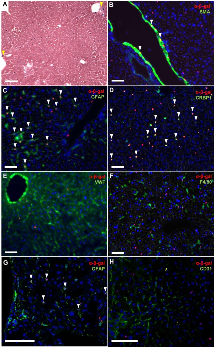
Figure 1. Hepatic Stellate Cells express RGS5.
A. Rgs5LacZ/LacZ mouse liver with X-gal labeling. RGS5+ peri-sinusoidal cells are distributed throughout the liver. RGS5 is also expressed in a subset of SMC of the portal vein (yellow arrows). B–G. Immunofluorescence (IF) for cell-specific markers in the Rgs5LacZ/LacZ liver. B. SMA and α-β-gal show RGS5 expression in vascular SMCs, as expected (arrows). C. GFAP and α-β-gal IF in Rgs5LacZ/LacZ mouse liver. β-gal+ nuclei are visible within GFAP+ astrocyte-like HSC cells, localizing RGS5 expression to HSCs (arrows). D. CRBP1 and α-β-gal IF are co-localized in HSC of Rgs5LacZ/LacZ liver (arrows). E. VWF (a marker of endothelial cells) and α-β-gal are not co-localized. VWF extends through all sinusoids, while β-gal+ cells are sparsely distributed. F. F4/80 (a maker of macrophage/Kupffer cells) and β-gal+ cells represent distinct cell populations. G. Confocal image of α-GFAP IF showing co-localization of the nuclear α-β-gal (arrows). H. Confocal image of CD31 (a marker of endothelial cells) and α-β-gal. CD31+ cells have β-gal− nuclei, while β-gal+ nuclei are not associated with CD31+ endothelial cells. All scale bars are 100 µm.

