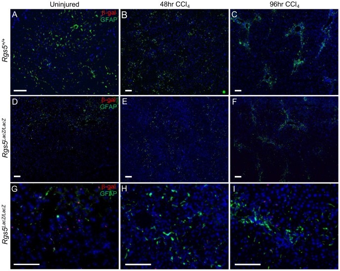
Figure 5. RGS5+ HSCs participate in the response to acute hepatic injury.
Anti-GFAP and anti-β-gal immunofluorescence were used to localize HSCs in acutely injured liver tissue. A–F. Low magnification images. G–I. High magnification of Rgs5LacZ/LacZ A,D Uninjured liver tissue from Rgs5+/+ and Rgs5LacZ/LacZ mice show sparse HSCs distributed throughout the liver. High magnification in uninjured Rgs5LacZ/LacZ shows HSCs are GFAP+ and β-gal+. B,E. At 48 hours post injury, HSCs are concentrated in the necrotic foci surrounding the central veins. β-gal+ cells (E,H) are associated with GFAP+ cells. C,F At 96 hours post injury, HSCs are tightly clustered at the foci of injury. I. β-gal+ cells are GFAP+. All scale bars are 100 µm.

