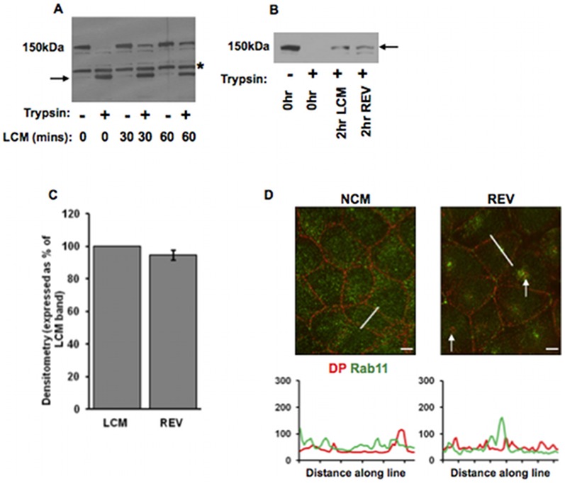
Figure 6. Internalised Dsg2 is not recycled to the cell surface.
(A) LCM-induced internalisation protects Dsg2 from trypsin. MDCK cells were treated with trypsin/EDTA after incubation LCM for 0, 30 or 60 minutes. Western blots show that cell surface Dsg2 (150 KDa) was degraded by trypsin/EDTA generating a membrane-protected cytoplasmic fragment (arrow) (LCM (mins) 0). (The band above the trypsin fragment and present in each lane is believed to be a natural degradation product of Dsg2.) However, after LCM treatment a substantial amount of full length Dsg2 remained after trypsin/EDTA treatment (LCM (mins) 30 and 60) showing that it had become membrane-protected. (B, C) The amount of membrane-protected Dsg2 (arrow) remains the same after induction of new desmosome formation following LCM treatment. Cells were treated either with LCM for 2 hours, or with LCM for 1 hour followed by NCM for 1 hour (total time 2 hours) to induce new desmosome formation (verified by immunofluorescence but not shown). The latter treatment is referred to as reverse calcium switching (REV). Western blots (B, quantified in C) show that the amount of membrane protected Dsg2 was identical after both treatments (2 hr LCM, 2 hr REV). Bar in C indicates s.e.m. (D) Internalised DP (arrows) does not colocalise with Rab11 following a reverse calcium switch (REV). Bar, 5 µm. Fluorescence profiles depict the intensity of staining along the white line in the images.

