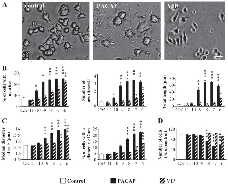
Fig. 1.
Effect of PACAP-38 and VIP on PC12 cell differentiation. A, microphotographs illustrating the neurite extension observed after 48 h of treatment with PACAP (100 nM) or VIP (100 nM). Scale bar, 15 μm. B, quantification of the percentage of cells with neurites, number of neurites per cell, and total neurite outgrowth after treatment with graded concentrations of PACAP or VIP (10 pM–1 μM). C, quantification of the median diameter of PC12 cells (micrometers) and the percentage of cells with a diameter above 17 μm after 48 h of treatment with graded concentrations of PACAP or VIP (10 pM–1 μM). D, quantification of the effect of graded concentrations of PACAP or VIP (10 pM–1 μM) on PC12 cell proliferation. * P < 0.05, **P < 0.01, *** P < 0.001 versus control.

