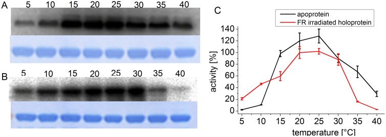
Figure 1. Autophosphorylation of Agp1 at various temperatures.
Autoradiogram (above) and Coomassie-stained blot (below) of Agp1 apoprotein (A) and FR irradiated holoprotein (B). The incubation temperature during the phosphorylation assay in °C is given above each lane. (C) Mean phosphorylation intensities of three experiments ± SE as shown in (A) and (B) are plotted over temperature. The 100% value corresponds to the mean signal of the holoprotein at 25°C.

