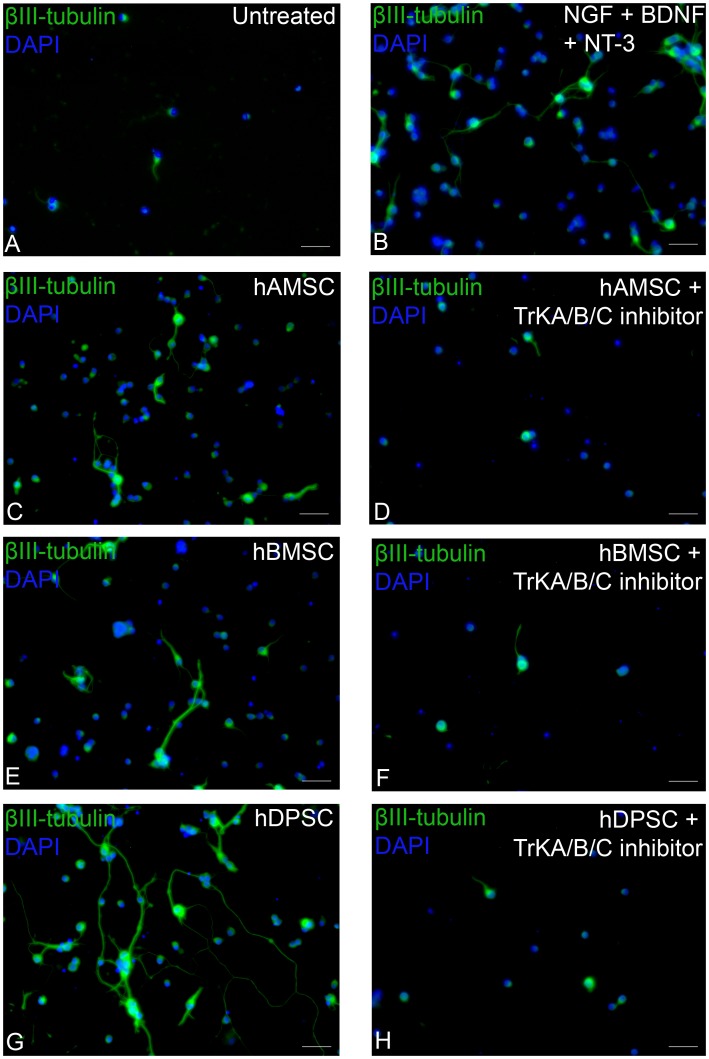
Figure 2. Immunocytochemical staining of RGC after retinal coculture with hDPSC, hBMSC and hAMSC in transwell inserts.
In vitro RGC cultured either alone (A), with exogenous neurotrophins (B), with hAMSC (with or without TrK inhibitors (C, D, respectively)), with hBMSC (with or without TrK inhibitors (E, F, respectively)) or with hDPSC (with or without TrK inhibitors (G, H, respectively). All images are representative of the entire culture, nine separate culture wells/treatment, with every three wells using a different animal (scale bars: 50 µm), cell nuclei were stained with DAPI.

