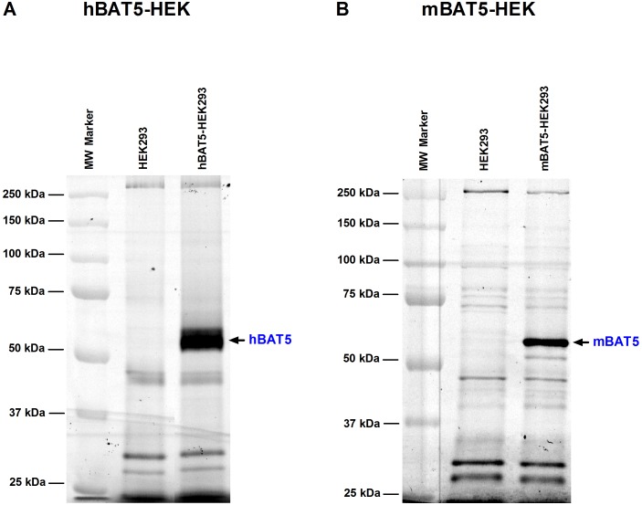
Figure 2. Activity-based protein profiling (ABPP) to visualize catalytically active BAT5 protein in lysates of HEK293 cells after transient expression of human (h) or mouse (m) BAT5 orthologs.
Serine hydrolases were labeled using the active site serine targeting fluorescent probe TAMRA-FP. After separation in SDS-electrophoresis gel (10%), serine hydrolase activity was visualized by in-gel fluorescent gel scanning as detailed in the Methods section. Molecular weight markers (MW) are indicated at left. Transient transfections with the cDNAs encoding hBAT5 (A) or mBAT5 (B) results in robust labeling of a ∼63 kDa protein band that is absent from parental HEK293 cells. Data are from one typical transfection, transfections were repeated twice with similar outcome.

