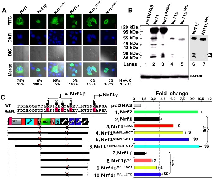
Figure 7. Blockage of Nrf1γ results an increase in the transactivation activity of Nrf1 and Nrf1β/LCR-F1.
(A) Confocal imaging of COS-1 cells that had been transfected with 1.3 µg DNA of each expression construct for Nrf1, Nrf1β and Nrf1γ or mutants, before their subcellular locations were then examined by immunocytochemistry with FITC-labelled second antibody in order to locate V5-tagged proteins. Nuclear DNA was stained by DAPI. The merge signal represents the results obtained when the two images were superimposed with DIC from normal light microscopy. Bar = 20 µm. The quantitative data (bottom) were calculated as described in Figure 2D. (B) Western blotting of COS-1 cells that had been transfected with the indicated expression constructs for V5-tagged Nrf1, Nrf1β and Nrf1γ and their point mutants (Met into Leu, below). The right panel shows that the same gel as the left panel was exposed to X-ray for a little longer time. Two bands representing the 36-kDa Nrf1γ and a 38-kDa polypeptide are indicated (arrows). GAPDH served as an internal control to verify the amount of proteins applied to each electrophoresis well. (C) Schematic representation of Nrf1, Nrf1β, and their Met-to-Leu mutants with various deletions. The upper left panel shows amino acids adjoining five numbered Met residues; their mRNA codons can be recognized by ribosome for the internal initiation to translate Nrf1β or Nrf1γ. The first four or all five Met-to-Leu mutants were made respectively to yield Nrf14xM/L and Nrf15xM/L, whilst Nrf1β M/L contains the fifth Met-to-Leu mutant. Additional deletion mutants were created on the base of Nrf15xM/L and Nrf1βM/L. The right panel shows luciferase reporter activity of COS-1 cells that had been transfected with 1.2 µg of each of indicated expression constructs, together with 0.6 µg of PSV40nqo1-ARE-Luc and 0.2 µg of β-gal plasmids. The data are shown graphically as fold changes (mean ± S.D.) of transactivation by indicated factors. Significant increases ($, p<0.05 and $$, p<0.001, n = 9) in the activity are compared to the activity of the intact Nrf1 or Nrf1β.

