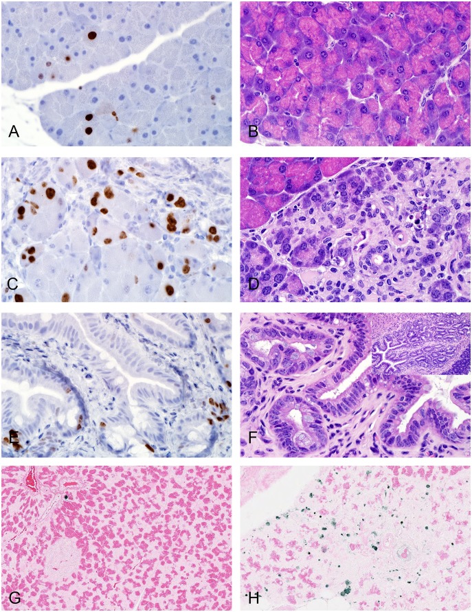
Figure 4. Representative micrographs comparing proliferative (Ki-67) and apoptosis (TUNEL) immunoreactivity to morphologic changes in HFD mice.
(A & B) Saline, 12 weeks, Ki-67 staining (A) was occasionally identified in nuclei in areas of normal acinar and centroacinar cells (B). (C & D) 30 µg/mg exenatide, 12 weeks, increased Ki-67 immunoreactivity (C) characterized by more frequently stained nuclei in acinar, centroacinar, ductal, and interstitial cells observed in areas of severe acinar cell injury (D) including autophagy, apoptosis, and necrosis and ductal metaplasia. Two nuclei placed side by side with basophilic stained cytoplasm are suggestive of cell proliferation (hyperplasia). (E & F) 30 µg/mg exenatide, 12 weeks, increased Ki-67 immunoreactivity (E) characterized by nuclear staining in epithelial cells of the main duct observed in areas of pseudostratified columnar epithelium with increased number of goblet cells (F). Inset in F showing main pancreatic ductal cell proliferation. (G) Saline, 12 weeks, TUNEL staining revealed an occasional acinar cell undergoing apoptosis. (H) 30 µg/mg exenatide, 12 weeks, revealed many apoptotic cells in areas associated with acinar cell injury. A–H, X600, A, C, E, (IHC for Ki-67); B, D, F (H&E stain); G & H (TUNEL). Inset in F, X200.

