
Figure 6. ERK pathway is not involved in ADP-induced cell proliferation inhibition.
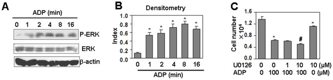
(A) ADP induced phosphorylation of ERK. HUVEC, starved overnight with serum-free medium, were treated with 100 µM ADP, and lysed at the indicated time points. Western blot was performed for the detection of phospho-ERK1/2 and total ERK1/2 respectively. β-actin protein was detected as a loading control. (B) Quantitation of phosphorylated ERK normalized to total ERK in (A). * P<0.05 compared with the control group. (C) The effect of ERK inhibitor, U0126, on ADP-induced cell proliferation inhibition. HUVEC, pre-treated with the indicated concentrations of U0126 for 30 min, were re-treated with the indicated concentrations of ADP for 24 h, followed with the same treatment once a day in the next 2 days. Cell number was measured by CCK-8 assay. * P<0.05 compared with the non-treated group. # P<0.05 compared with ADP-treated alone group.