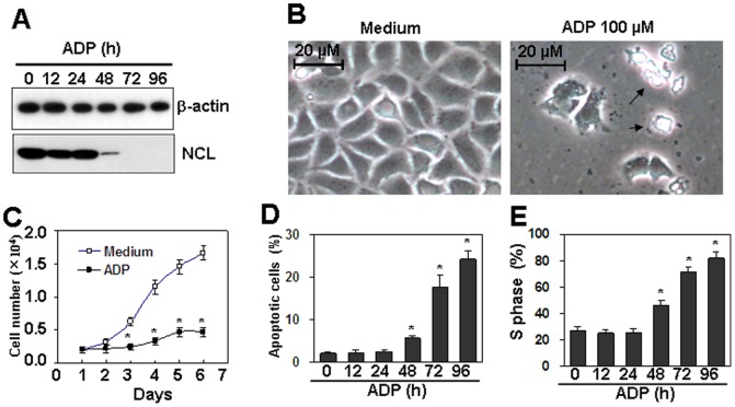
Figure 9. The effect of ADP on the proliferation of cervical cancer cells.
(A) ADP down-regulated the protein levels of nucleolin in Caski cervical cancer cells. Cells were treated with 100 µM ADP for the indicated time periods. Nucleolin protein levels were detected by western blot. β-actin protein levels were detected as loading controls. (B) Microscope observation of Caski cells treated with ADP. Cells were treated with 100 µM ADP for 72 h. Cell morphology in the presented field was obtained by microscope. Arrows indicated the detached cell debris. (C) ADP inhibited proliferation of cervical cancer cells. Caski cells were treated with 100 µM ADP for the indicated time periods. Cell numbers were detected every day by CCK-8 assay. P<0.05 compared with the control group. (D) ADP induced cell apoptosis in cervical cancer cells. Caski cells were treated with 100 µM ADP for the indicated time periods. Cell apoptosis was measured by PI/FITC-Annexin V staining assay. P<0.05 compared with the control group. (E) ADP induced cell cycle arrest in S phase. Caski cells were treated with 100 µM ADP for the indicated time periods. Cell cycle was measured by PI staining. P<0.05 compared with the control group.

