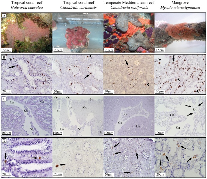
Figure 2. Cell proliferation and cell loss in a selection of four sponge species.
Sponge species from three benthic ecosystems; tropical coral reef, temperate Mediterranean reef, and mangroves. (A) In situ (H. caerulea, C. reniformis and M. microsigmatosa) and ex situ (C. caribensis) photographs of test species. (B) BrdU-positive choanocytes (arrows) and mesohyl cells (arrowheads) of sponges BrdU-labeled for 6 h (10 h for C. reniformis) in vivo as a measure for proliferation. Areas of non-specific BrdU-labeling are occasionally seen in the cytoplasm of cells or extracellularly. (C) High amounts of cell shedding (Sh) in the lumen of excurrent canals (Ca) in specimens of H. caerulea, C. caribensis and C. reniformis sampled in situ. Choanocyte chambers (Ch), oscula (Os), the mesohyl (Me) and pinacoderm (Pi) are shown. Minor amounts of cell shedding (arrows) in the tropical mangrove sponge M. microsigmatosa sampled in situ. (D) Active caspase-3 activity of in vivo tissue was found in cells located in the mesohyl (arrows) resembling spherulous cells and, occasionally, archeocytes.

