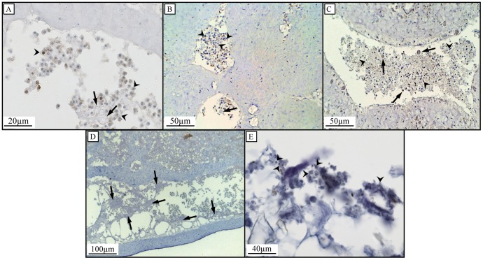
Figure 3. Histological investigations of cell shedding and identification of cellular debris in sponge-derived detritus.
Arrowheads indicate shed choanocytes and arrows indicate shed spherulous cells in H. caerulea (A), C. caribensis (B) and C. reniformis (C). Shed cells in H. caerulea are present as mucal sheets (arrows) of cellular debris when close to the outflow openings (D). Light microscopy of detritus samples shows the presence of degraded cellular material, indicated by arrowheads (E).

