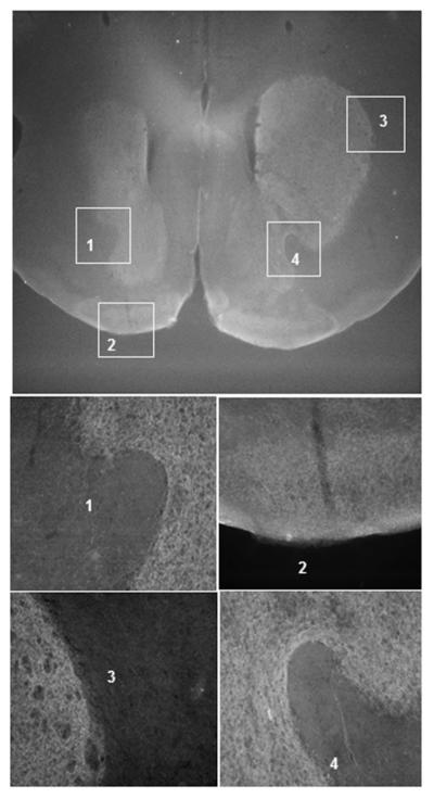
Figure 4. Fluorescence immunostaining of the AAV mediated wild type DAT expression in DA terminal brain regions of DAT-CI mice injected with AAV-DATwt in the ventral midbrain.

Coronal brain slices from DAT-CI mice with AAV vector injected in vMB. The brain slices were stained with anti HA tag primary antibody and goat anti mouse Cy3 secondary antibody. The rostral/caudal coordinate for the image is about +1 mm relative to Bregma. Images 1- 4 are higher magnification images of the boxed regions specified.
