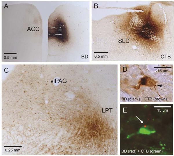
Figure 5.
The projections of the mPFC to REM sleep circuit. To investigate a possible pathway by which the dmPFC may be modulating REM sleep, two tracers were injected (both unilaterally) into an individual animal: A BD, an anterograde tracer, into the dmPFC (Bregma 3.5mm); B CTB, a retrograde tracer, into the REM-on SLD (Bregma −9.4mm). C Cells in the vlPAG-LPT (Bregma −7.2mm) were then sought that were stained for CTB (brown) and also had BD boutons (black) (D). E. A series from the same case was stained with CTB (AlexaFluor488 green) and BD (Cy3 red) and viewed under a confocal microscope (63x). A cell in the vlPAG is stained green, indicating it projects to the SLD area, and has a red bouton from the vmPFC (arrow). This image was taken in a single optical plane.

