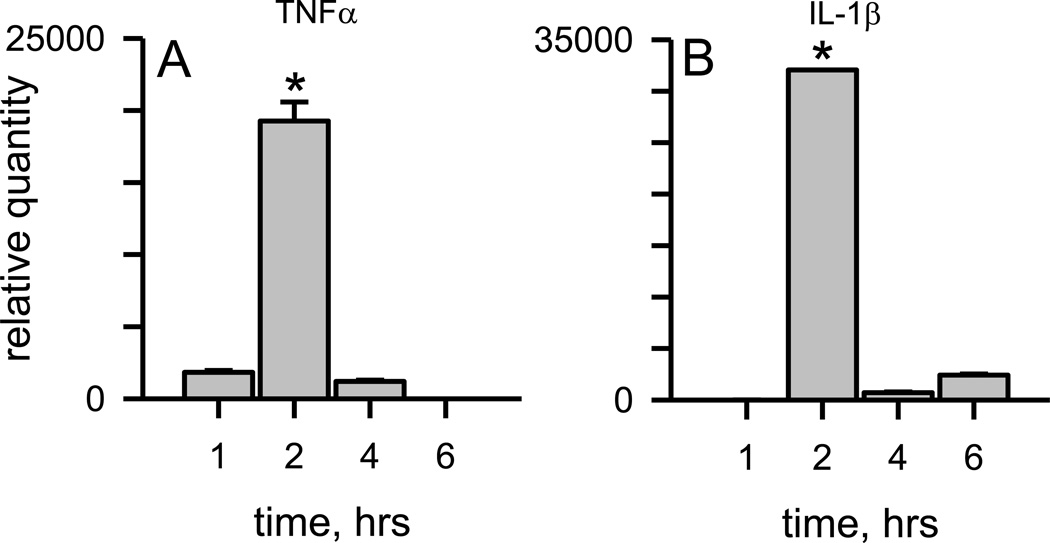
Figure 1.
Aβ(1 −42) protofibrils formed and isolated in aCSF are significant stimulators of TNFα and IL-1β transcription. SEC-isolated Aβ(1–42) protofibrils in aCSF were incubated with WT primary microglia at a final concentration of 1 5 µM for 1, 2, 4, and 6 hrs in serum-free medium. At each time point the cells were lysed for total RNA extraction. TNFα (Panel A) and IL-1β (Panel A) mRNA levels were measured by qPCR as described in the Methods. The term relative quantity is described in the Methods and represents the mRNA comparison between microglia treated with Aβ(1–42) protofibrils and those treated with buffer control. β-actin was used as an internal control in separate experiments and did not vary significantly between samples. Data bars represent the mean ± std error of n=8 qPCR replicates. TNFα and IL-1β mRNA levels at 2 hours were significantly different than any other time point (*p<0.001).

