Figure 4.
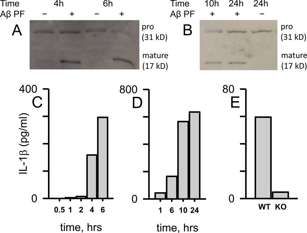
Aβ(1–42) protofibrils stimulate time- and MyD88-dependent intracellular mature IL-1β accumulation in microglia. Panel A, C. WT primary microglia were treated with SEC-isolated Aβ(1–42) protofibrils (15 µM) in aCSF (+) or just aCSF buffer (-) and incubated for 0.5, 1, 2, 4, and 6 hrs in serum-free medium at 37 °C. At each time point the cells were lysed and extracts prepared for intracellular IL-1β Western blot (Panel A) and ELISA (Panel C) analysis. A separate but similar experiment was done over a longer time course with the same analysis (Panel B Western blot, Panel D ELISA). Each intracellular cell extract sample for all panels was obtained from a combination of five replicate wells in a 96-well plate treated with 20 µL total of lysis buffer, thus the band or data bar is representative of a 5-well extract pool. Panel E. Intracellular mature IL-1β was measured by ELISA in extracts prepared as described above after treatment of WT and MyD88−/− primary microglia (KO) for 6 hrs with Aβ(1–42) protofibrils (15 µM) as described in Panel A and C. Corresponding control treatments with a volume of aCSF equal to that carrying the Aβ were also analyzed for IL-1β. These control levels, which averaged 10% of the Aβ-stimulated levels, were subtracted from the overall Aβ response. The measured intracellular IL-1β levels in C-E (pg/ml), which were much higher, have been normalized to reflect the response from pooling of multiple wells containing 104 microglial cells per well and in a lesser volume.

