Figure 5.
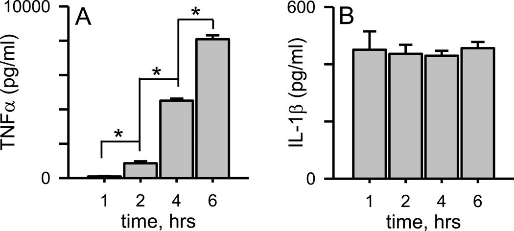
Aβ(1–42) protofibrils stimulate rapid IL-1β secretion but a slower time-dependent TNFα secretion. SEC-isolated Aβ(1–42) protofibrils in aCSF were incubated with WT primary microglia at a final concentration of 15 µM for 1, 2, 4, and 6 hrs in serum-free medium. At each time point the conditioned microglial medium was collected for TNFα and IL-1β protein determination by ELISA. Panel A. TNFα protein levels were determined from the supernatant of individually treated wells. Data bars represent the mean ± std error of n=5 replicates at each time point. Control treatments with an equal volume of aCSF produced very low TNFα levels compared to protofibrils ranging from 36–290 pg/ml (<4% of the Aβ-stimulated response) at the different time points and were subtracted from Aβ-stimulated samples. TNFα levels at each successive time point are statistically different than the preceding time point (p<0.001). Panel B. IL-1β protein levels were measured in the same manner as for TNFα. Control treatments with an equal volume of aCSF produced IL-1β levels of 1 pg/ml and were subtracted from Aβ-stimulated samples. The length of incubation time had no statistical difference on the amount of secreted IL-1β (p>0.05).

