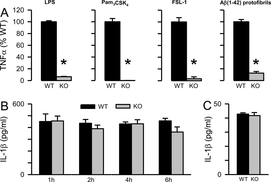
Figure 6.
Aβ(1–42) protofibril-induced TNFα secretion, but not rapid IL-1β secretion, is dependent on MyD88. Panel A. TLR4 ligand LPS, TLR1/2 ligand Pam3CSK4, TLR2/6 ligand FSL-1, and SEC-isolated Aβ(1–42) protofibrils were incubated with WT and MyD88−/− (KO) primary microglia at a final concentration of 100 ng/ml for all TLR ligands and 15 µM for Aβ for 6 hrs in serum-free medium. Secreted TNFα was measured by ELISA in the conditioned medium. Data bars represent the average ± std error of n=9 replicates (triplicates from 3 separate experiments) for LPS, Pam3CSK4, and FSL-1, and n=6 replicates (triplicates from 2 separate experiments) for Aβ(1–42) protofibrils. Data is presented as % WT response. Actual TNFα levels were 21667, 4584, 2888, and 12652 pg/ml for LPS, Pam3CSK4, FSL-1, and Aβ(1–42) protofibrils respectively. Control treatments with an equal volume of H2O or aCSF were less than 3% of the TNFα response and were subtracted from TLR ligand- or Aβ-stimulated samples. Statistical analysis showed a significant difference (p<0.005) between the WT and MyD88−/− results for all four treatments. Panel B. SEC-isolated Aβ(1–42) protofibrils were incubated with WT primary microglia and MyD88−/− (KO) microglia at a final concentration of 15 µM for 1, 2, 4, and 6 hrs in serum-free medium. Secreted IL-1β was measured by ELISA in the conditioned medium. Data bars represent the average ± std error of n=5 replicates. Control treatments with an equal volume of aCSF produced 1 pg/ml IL-1β at all time points for both the WT microglia and the MyD88−/− microglia and were subtracted from Aβ-stimulated samples. Statistical analysis showed no significant difference between the WT and MyD88−/− results at any time point (p>0.05). Panel C. Secreted IL-1β was measured by ELISA after treatment of WT and MyD88−/− primary microglia (KO) with Aβ(1–42) protofibrils (15 µM) for 6 hrs in serum-free medium. Conditioned medium was collected and secreted IL-1β was measured by ELISA. The secreted data bars are the average ± std error for n=5 replicates. Control treatments with an equal volume of aCSF were subtracted from Aβ-stimulated samples and averaged 0.4%. No statistical difference was observed between the WT and MyD88−/− response (p>0.05).

