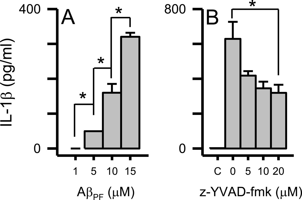
Figure 7.
Dose-dependent IL-1β secretion in primary microglia induced by Aβ(1–42) protofibrils. Panel A. Primary microglia were treated with SEC-isolated Aβ(1–42) protofibrils (1, 5, 10, and 15 µM) and incubated for 6 hrs. Secreted IL-1β levels were measured in the conditioned medium by ELISA. Data bars represent the average ± std error of n=3 replicates for each condition. Statistical differences (p<0.05) in secreted IL-1β elicited by each Aβ protofibril concentration are denoted with an asterisk. Panel B. Primary microglia were pretreated as described in the Methods with increasing concentrations of the caspase-1 inhibitor z-YVAD-fmk or 0.5% DMSO vehicle followed by 15 µM Aβ(1–42) protofibrils. A control treatment (C) that contained 0.5% DMSO but neither z-YVAD-fmk or Aβ was also included. Secreted IL-1β levels were measured in the conditioned medium by ELISA. Data bars represent the average ± std error of n=3 replicates for each condition. Statistical differences (p<0.05) in secreted IL-1β elicited by Aβ protofibrils in the absence or presence of the inhibitor are denoted with an asterisk.

