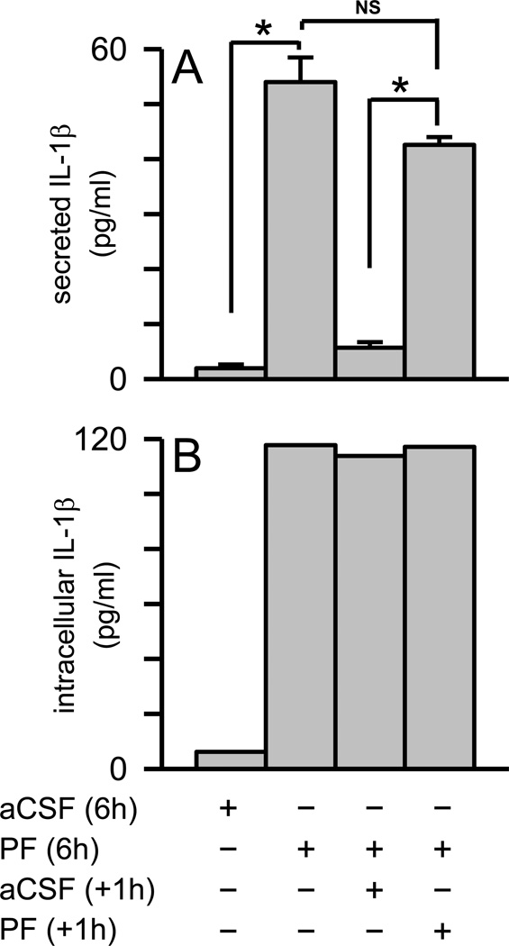
Figure 9.
A second stimulus of microglia with Aβ protofibrils secretion does not release accumulated intracellular IL-1β . Primary microglia were exposed to SEC-isolated Aβ(1–42) protofibrils (15 µM) or aCSF buffer alone and allowed to incubate at 37 °C for 6 hrs. The conditioned medium was then removed and one intracellular extract was prepared for each condition (combination of five wells in 20 µL of lysis buffer). The measured intracellular IL-1β response in pg/mL, which was much higher, was normalized based on the number of wells and lysis buffer volume. For the remaining cells, fresh medium was applied along with a second protofibril (n=5 wells or replicates) or buffer treatment (n=5 replicates). After an additional 1 hr incubation, the conditioned medium from each well was removed separately and one cell extract was again prepared for each condition. Secreted (Panel A) and intracellular (panel B) IL-1β was determined by ELISA. IL-1β data bars represent the average ± std error of n=5 replicates for secreted and one intracellular extract for each condition. Statistically significant differences between treatments are denoted with an asterisk (p<0.001). No statistical difference (NS) was found between the microglial IL-1β response when treated with Aβ(1–42) protofibrils for 6 hr and again for 1 hr (p>0.05).

