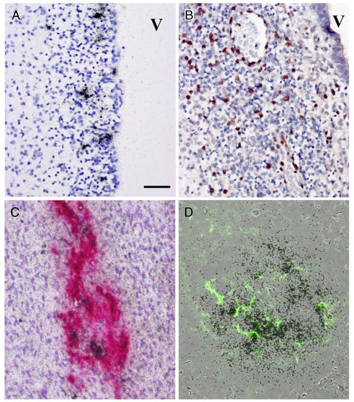
Fig. 2.
Histopathological assessment of brain, adrenal gland and spleen. Histopathological analysis of brain tissues demonstrates widespread inflammation and infection in germinal matrix (A,B). In situ hybridization (ISH) for coxsackie B virus (CB4) (A) shows infection of stem cell elements underlying ependymal lined ventricle (V). Immunocytochemistry for CD3 demonstrates diffuse infiltration of germinal matrix by T cells (B). Double-label immunocytochemistry and ISH for tyrosine hydroxylase (red) and CB4 RNA (black grains) of the adrenal medulla shows tyrosine hydroxylase positive cells, some of which co-label for viral RNA (C). Overlain images of ISH for viral RNA in spleen demonstrating abundant signal (black grains) in lymphoid follicles while a serial paraffin section defines CD21 staining (green) of follicular dendritic cells (D). All images at 20× magnification, bar = 50 μm.

