
Figure 1.
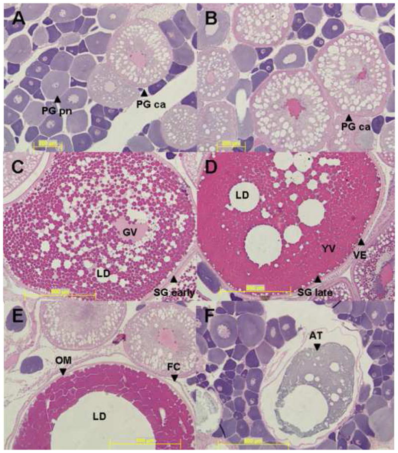
Representative micrographs of female largemouth bass ovarian stages. Female stages are as follows; A) Primary growth perinuclear (PG pn); B) primary growth cortical alveoli (PG ca); C) Secondary growth early (Sg early); D) Secondary growth late (Sg late); E) Oocyte maturation (OM); and F) Atresia (AT). Additional abbreviations are as follows: Follicle Cells (FC), Germinal Vesicle (GV), Lipid Droplet (LD), Vitelline Envelope (VE), and Yolk Vesicles (YV). Scale bars correspond to 200 μm (A–B) and 500 μm (C–F).