
Figure 4.
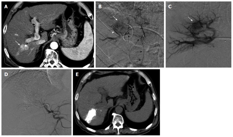
Transportal chemoembolization for hepatocellular carcinoma with an arterioportal shunt in a 70-year-old man. Enhanced abdominal computed tomography (CT) examination (A) demonstrated a recurrent hepatocellular carcinoma (HCC) (A, arrow) in the posterior segment with a lipiodol deposit (arrowhead). Hepatic arteriography via the anterior branch (B) revealed a HCC (B, white arrow) fed by the cystic artery (B, black arrows) and an intratumoral arterioportal shunt (B, arrowhead). A 5-French sheath was inserted into the portal vein via the lateral superior branch of the left portal vein. Direct portography via the portal vein branch contributed an arterioportal shunt showed a tumor stain (C, arrow). Following transcatheter portal chemoembolization, proper hepatic arteriography showed non-visualized tumor stain and arterioportal shunt (D). Non-enhanced CT 7 d after therapy (E) showed a dense accumulation of lipiodol in the tumor and peritumoral liver parenchyma.