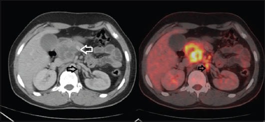
Figure 3.

Left Image: Mass with hypodense necrotic areas in head of pancreas. It is seen closely abutting the superior mesenteric vein (white arrow). Small paraaortic lymph node is also seen (black arrow). Right image: corresponding positron emission tomography image showing intense fluorodeoxyglucose uptake
