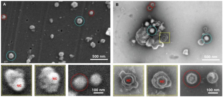
Figure 4.
Imaging herpes simplex virus. SEM (A) and TEM (B) images of the herpes virus are presented, showing intact virus (blue circle), partially disrupted (yellow squares), and nucleocapsids (red circles). In this case both methods detected the virus, however the staining in the TEM images provide a superior diagnostic identification, for example the nucleocapsid (NC) has superior staining in the TEM sample preparation.

