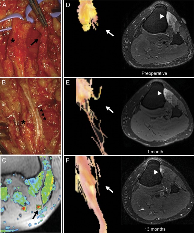
Figure. Magnetic resonance tractography demonstrating axonal regeneration.
Operative exploration of the left peroneal nerve identified a damaged, scarred deep peroneal nerve branch (black arrow, A) and relatively preserved superficial peroneal nerve branch (asterisk, A and B). The deep peroneal nerve branch was resected and the resulting defect bridged with 3 sural nerve grafts (black arrowheads, B). A thresholded fractional anisotropy (FA) map (C) is shown fused to the corresponding axial T1-weighted image. The proximal common peroneal nerve is identified as a region of increased FA signal (arrow), and is encircled by a region of interest used to calculate average FA values. Preoperative magnetic resonance (MR) tractography demonstrates the proximal stump of the deep peroneal nerve branch with no axons identified distal to the point of transection (white arrow, D), and T2-weighted, fat-suppressed imaging of the proximal left leg identifies denervation edema in deep-peroneal innervated muscles (tibialis anterior and extensor digitorum longus, white arrowhead). Repeat MR tractography performed 1 month after surgical repair identifies sparse, disorganized axons distal to the proximal graft neurorrhaphy (white arrow), and persistent muscle edema (E). Tractography performed 13 months after nerve graft repair demonstrates an organized bundle of fibers traversing the nerve grafts and resolution of muscle edema (F).

