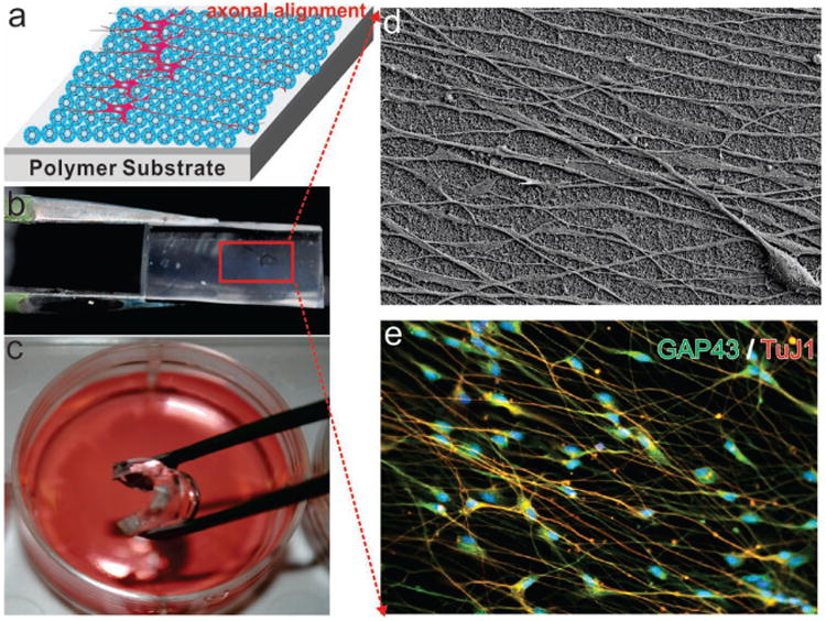
Figure 5.

Axonal Alignment of differentiated hNSCs on SiNP-GO on flexible and biocompatible substrates made from polydimethylsiloane (PDMS). (a) schematic diagram of axonal alignment of differentiated hNSCs on SiNP-GO on polymer substrates. b) SiNP-GO monolayer on PDMS. c) Flexible PDMS substrate with SiNP-GO in media for culturing hNSCs. d) SEM image of SiNP-GO on PDMS substrate showing highly aligned axons from hNSCs on Day 14. e) Immunocytochemistry results showing the expression of neuronal marker TuJ1 and axonal marker GAP43 in hNSCs.
