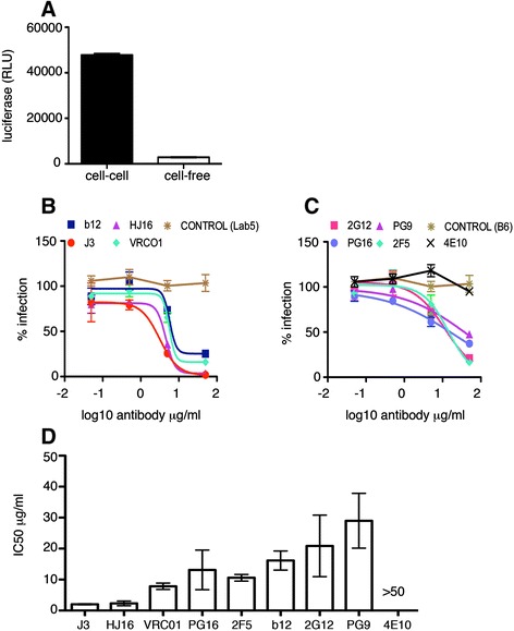
Figure 1.

Inhibition of HIV-1 T cell-T cell spread by anti-CD4 binding site, MPER and glycan-specific antibodies. (A) Quantification of the luciferase signal in Jurkat 1G5 cells contributed by cell-cell and cell-free spread as described in the methods. Data show the mean and SEM from 3 independent experiments. (B) Antibodies targeting the CD4 binding site, (C) the gp41 MPER and gp120 glycans and non neutralising antibody controls (B6 and Lab5), were serially diluted and incubated with HIV-1 (NL4.3) infected Jurkat T cells for 1 h at 37°C. Uninfected target T cells containing a luciferase-reporter gene were added and cells incubated for 24 h as described in the methods to allow for cell-cell spread of HIV-1. Data are shown as the percentage neutralisation normalised to virus-only controls and representative of three independent experiments. (D) The average IC50 values (μg/ml) were generated from duplicate titrations of the indicated antibodies in three independent experiments and show the mean with the SEM.
