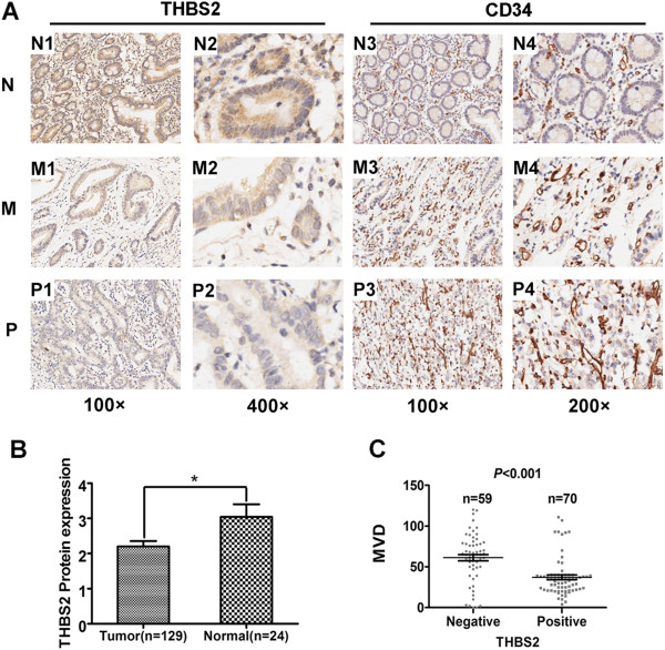
Figure 2.

Immunohistochemical analysis of THBS2, CD34. (A) Representative images of THBS2 and CD34 expression: N1, N2, N3, N4, normal gastric tissue (N); M1, M2, M3, M4, moderately differentiated (M); P1, P2, P3, P4, poorly differentiated (P). Magnification: 100× (N1, M1, P1, N3, M3, P3), 200× (N4, M4, P4) and 400× (N2, M2, P2). (B) Comparison of THBS2 expression of gastric cancer (n = 129) and normal gastric tissues (n = 24) in TMA. The THBS2 expression level presented as mean ± SEM. (C) Comparison of MVD in THBS2-positive gastric cancer group (n = 70) and THBS2-negative gastric cancer group (n = 59). Dots represent MVD of each samples. Values are means ± SEM.
