Figure 3.

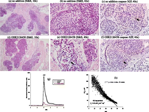
Breast lobular carcinoma in situ. (a-f) Histology and expression of caspase-3 by immunoperoxidase. (a-b) Untreated tumor demonstrating expanded acini by an in situ proliferation of uniform neoplastic cells, H&E at 10× and 40×, respectively. (d-e) Treated in situ carcinoma demonstrating fragmentation and degeneration of neoplastic cells (black arrow), H&E at 10× and 40×, respectively. (c and f) Untreated and treated tumor showed rare positivity (1%, black arrow) for caspase-3, immunoperoxidase (IP), 40×. (g) HPLC runs of intracellular caspase-3 activity in treated and untreated tumor. The AMC peak (retention time, ~4.8 min) in treated tumor was blocked by zVAD. (h) Cellular respiration, measured immediately on tumor arrival to the laboratory to affirm viability.
