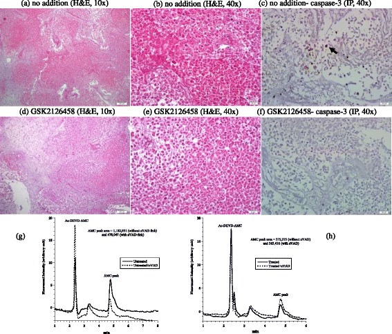
Figure 7.

Metastatic colorectal adenocarcinoma. (a - f) Histology and expression of caspase-3 by immunoperoxidase. (a - b and c - d) Sample consisted of necrotic neoplastic tissue, H&E, 10× and 40×, respectively. (c and f) Caspase-3 demonstrated a positive staining (black arrow) in up to 5% of necrotic neoplastic cells in untreated tissue and absence of staining in treated tissue, immunoperoxidase (IP), 40×. (g - h) HPLC runs of intracellular caspase-3 activity in treated and untreated tumor (AMC peak retention time, ~4.8 min).
