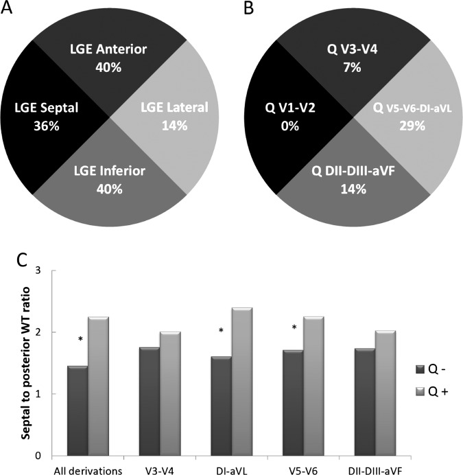
Figure 2.
Illustration of discordance between location of Q waves on ECG derivations and location of LGE in the myocardium on CMR. (A) Incidence of LGE within cited location among all patients (n=42). LGE was predominantly on the anterior, septal and inferior territory. (B) Incidence of Q waves in the cited derivations among all patients. Q waves were more likely in the derivations corresponding to the lateral territory. (C) Comparison of septal to posterior WT ratio according to the presence (Q+) or absence (Q−) of Abnormal Q waves in their different derivations. * p<0.05. CMR, cardiac MRI; HCM, hypertrophic cardiomyopathy; LGE, late gadolinium enhancement; WT, wall thickness.

