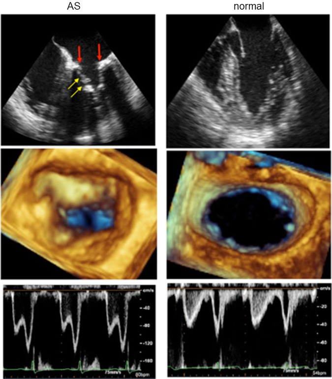
Figure 1.
Two-dimensional (2D) and Three-dimensional (3D) images of the mitral valve and transmitral flow profile in a patient with AS and a control participant. On the 2D image, mitral annular calcification (red arrow) was prominent in the patient with AS. The 3D zoomed image from a left atrial perspective showed a whole circumferential mitral annular calcification resembling a prosthetic mitral valve ring. In this patient with AS, the leaflet opening was also restricted (yellow arrow) and the peak transmitral flow velocity increased to 160 cm/s.

