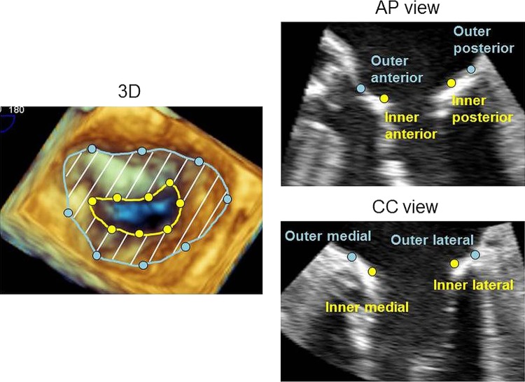
Figure 2.
Three-dimensional (3D) measurement of the outer and inner/effective mitral annular area. The left panel showing a cropped 3D image of the mitral valve. From this 3D data set, an apical long-axis view of the mitral apparatus was used to determine the anterior and posterior mitral annulus, which was perpendicular to the commissure–commissure view and crossed the centre of the commissure–commissure plane (medial–lateral). The outer and inner borders of the annulus were then manually traced in multiple planes rotated around the axis connecting the centre of the mitral annulus and left ventricular apex, to measure the outer and inner/effective mitral annular area.

