Fig. 2.

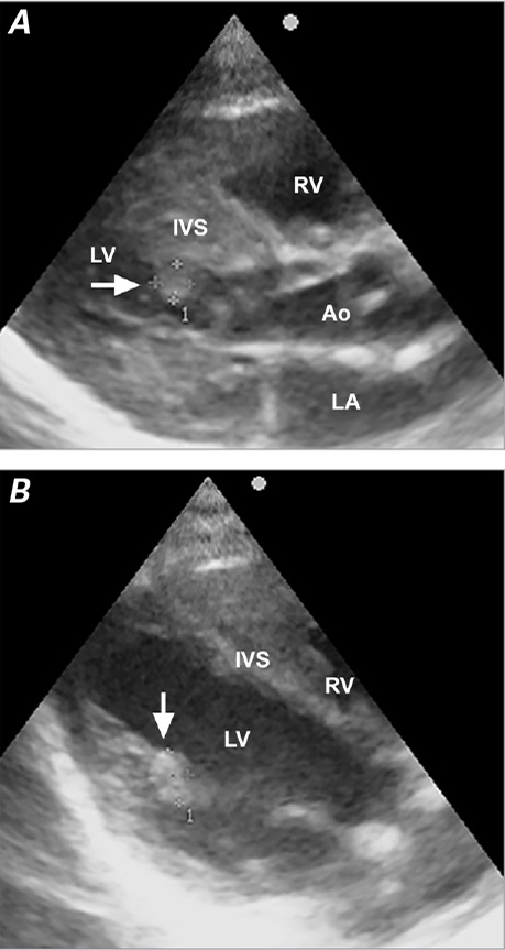
Echocardiograms show rhabdomyomas attached to the A) interventricular septum (arrow) and B) papillary muscle of the mitral valve (arrow).
Ao = aorta; IVS = interventricular septum; LA = left atrium; LV = left ventricle; RV = right ventricle

Echocardiograms show rhabdomyomas attached to the A) interventricular septum (arrow) and B) papillary muscle of the mitral valve (arrow).
Ao = aorta; IVS = interventricular septum; LA = left atrium; LV = left ventricle; RV = right ventricle