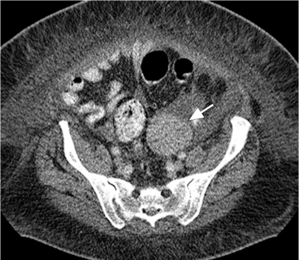
Fig. 3.

Computed tomogram of the abdomen and pelvis shows the ovarian tumor, a 6 × 6-cm left adnexal mass (arrow), medial to the left iliac process, with well-defined margins. The homogeneous mass displays hypercontrast enhancement without fat or calcification. No invasion of surrounding structures or abnormal lymph node in the pelvis suggests any metastatic disease.
