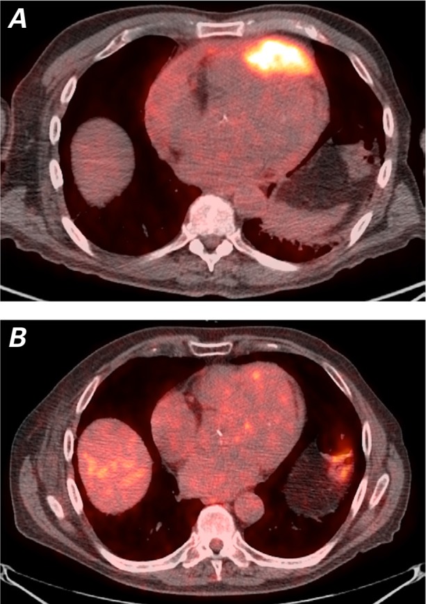
Fig. 6.

Positron-emission tomographic/computed tomographic images (axial views) show A) the large mass in the anterior portion of the heart at the time of presentation and B) substantial reduction of tumor size post-radiotherapy. Almost no tracer uptake is visible at the site of the previously existing hypermetabolic mass.
