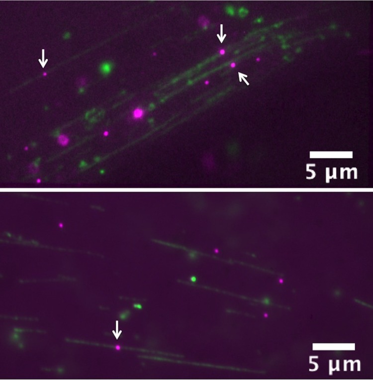
FIG. 4.
λ-phage DNA (green)—E. coli RNAP complexes (magenta) combed on LP O2 plasma treated PSQ surface. White arrows indicate the candidate DNA-RNAP complexes. DNA molecules were labeled with YOYO-1 nucleic acid stain (1/5 of dye/base-pair ratio) and RNAP with primary-secondary antibody coupling scheme.28 Some QDs also non-specifically immobilized on the surface (bottom), but false positives can be identified using electric-field assisted stretching/recoiling of DNA-protein complexes. (Multimedia view) [URL: http://dx.doi.org/10.1063/1.4892515.5]

