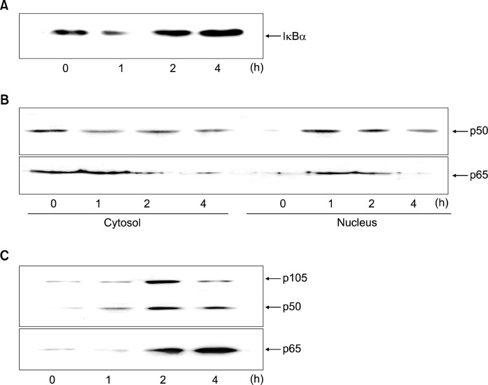
Fig. 1.
Protein levels of IκBα, p105, p50 and p65 in H. pylori-infected AGS cells. The cells were seeded in 12-well culture plates at 105 cells per well and cultured to reach 80% confluency. The bacterial cells were added to the cultured cells at a bacterium/cell ratio of 500:1. The cells were cultured for 4 h. The protein levels in whole cell extracts (A, C) and the protein levels in cytosolic extracts and nuclear extracts (B) were shown. Western bot result in each lane is the representative of five separate experiments.

